
Chronic Venous Insufficiency (CVI)- A Silent Epidemic, Its Hidden Complications, and the Dangers of Social Media Hype
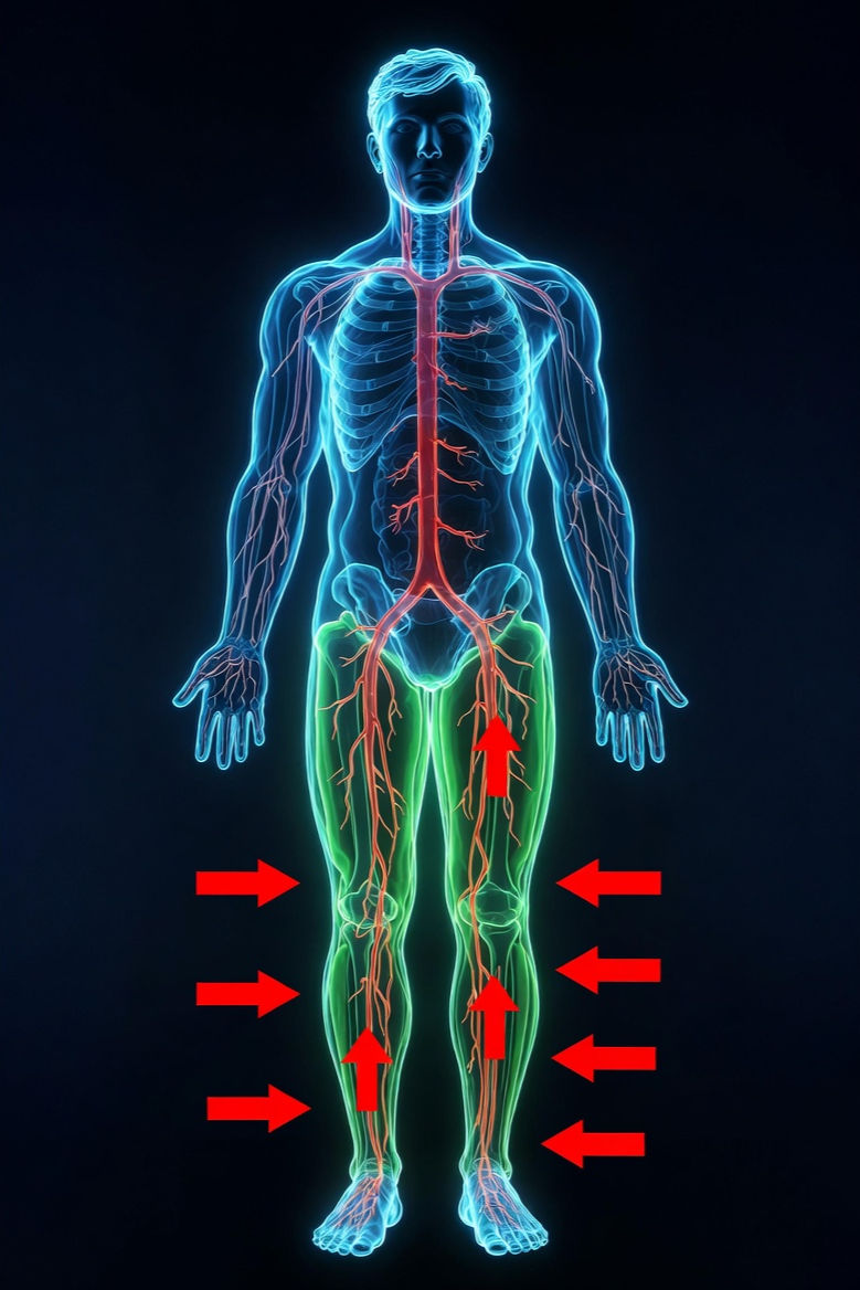
Chronic venous insufficiency (CVI) affects millions worldwide and often goes unnoticed until it leads to more serious complications. CVI is one of the most common yet underappreciated vascular conditions affecting millions worldwide. Often dismissed as simple “varicose veins”, “tired legs" or ”lymphatic sluggishness". When the veins in the legs fail to efficiently return blood to the heart, pressure builds up in the veins. This leads to venous hypertension, pooling, inflammation, and a cascade of symptoms that can severely impact quality of life. This pressure can cause swelling, skin changes, and eventually venous stasis dermatitis and skin breakdown and a host of other issues. Understanding how CVI progresses and recognizing early signs can help manage symptoms and may help prevent severe damage.
What Is Chronic Venous Insufficiency?
CVI arises primarily from incompetent venous valves, vein wall weakness, or obstruction (often post-thrombotic). These valves normally prevent blood from flowing backward. When they fail, blood pools in the lower legs, increasing venous pressure causing ambulatory venous hypertension.
Pathophysiology and Progression: From Mild Symptoms to Severe Disease
This progression leads to capillary leakage, inflammation, hemosiderin deposition (brownish skin staining), lipodermatosclerosis (hardened skin), and eventually tissue breakdown.. This condition is common in adults over 50, especially those who stand or sit for long periods, are overweight, or have a history of deep vein thrombosis (DVT).
Symptoms of CVI
Symptoms start subtly:
Leg swelling, especially after prolonged sitting or standing
Aching, heaviness, or cramping in the legs
Visible varicose veins
Skin discoloration or thickening around the ankles
If untreated, CVI can worsen and lead to skin complications. Without intervention, progression follows the CEAP stages:
C1–C2: Telangiectasias, reticular veins, varicose veins.
C3: Edema.
C4: Skin changes (pigmentation, eczema).
C5–C6: Healed or active venous ulcers.
Secondary Lymphatic Involvement and Other Complications: The Underrecognized Cascade
One of the most critical yet frequently overlooked aspects of CVI is its impact on the lymphatic system, leading to phlebolymphedema (also called lymphedema of venous etiology or LOVE). This mixed venous-lymphatic insufficiency occurs when chronic venous hypertension overwhelms lymphatic drainage capacity.
How it develops: Excess fluid, proteins, and inflammatory mediators leak into tissues due to venous pressure. Lymphatics initially compensate but eventually become damaged or fibrotic from sustained overload, scarring, and inflammation. This creates a vicious cycle of worsening edema, fibrosis, and impaired healing. Phlebolymphedema is considered the most common form of secondary lymphedema in Western countries.
Consequences:
More pronounced, protein-rich swelling that responds less well to simple elevation.
Increased risk of recurrent cellulitis and infections.
Harder, thickened skin (fibrosis).
Greater difficulty healing ulcers.
Higher healthcare utilization and costs.
Studies emphasize that venous leg ulcers almost always involve some degree of lymphatic dysfunction. Early recognition and combined veno-lymphatic management (e.g., Complete Decongestive Therapy including Manual Lymphatic Drainage) are essential, as standard compression alone may be insufficient in mixed cases.
Other complications include:
Venous stasis dermatitis and recurrent skin infections.
Lipodermatosclerosis and atrophie blanche (painful white scars).
Bleeding from fragile varicosities.
Superficial thrombophlebitis or progression to deep vein issues.
Reduced mobility, depression, and social isolation.
Rarely, malignant transformation in long-standing ulcers (Marjolin’s ulcer).
Obesity and sedentary behavior exacerbate all these issues by increasing abdominal pressure, weakening the calf muscle pump, and promoting further inflammation.
How CVI Leads to Venous Stasis Dermatitis
Venous stasis dermatitis is an inflammatory skin condition caused by poor blood flow and fluid buildup in the lower legs. The increased pressure damages small blood vessels, causing red, itchy, and scaly skin. Over time, the skin becomes fragile and prone to injury.
Signs of Venous Stasis Dermatitis
Redness and inflammation around the ankles and lower legs
Itching and burning sensations
Dry, flaky, or scaly skin patches
Brownish discoloration due to iron deposits from leaking blood cells
This stage signals that CVI is progressing and requires medical attention to prevent further damage.
The False and Misleading Social Media Hype
Social media platforms are flooded with oversimplified “cures,” miracle supplements, essential oils, restrictive diets, or unproven devices promising to eliminate varicose veins or CVI overnight. These claims often lack rigorous evidence and can delay proper care.
Common myths and hype:
“Crossing legs or standing causes varicose veins” — association exists with prolonged standing/sitting, but crossing legs is not a primary cause.
“Only older women get it” — affects both sexes and all ages, though risk increases with age and pregnancy.
“Natural remedies (specific herbs, creams, or detoxes) cure CVI” — some adjuncts like horse chestnut or flavonoids may help symptoms mildly, but they do not replace compression or address underlying reflux.
“Surgery is always painful and outdated” — modern endovenous procedures are minimally invasive.
“Compression is unnecessary if you use this gadget/pill” — unsupported.
Quick-fix promises ignoring the chronic nature of the disease.
Such hype creates false hope, leads to wasted money on ineffective products, and can result in worsening disease when patients postpone evidence-based evaluation (duplex ultrasound, vascular specialist consultation). Regulatory bodies and medical societies stress that accurate diagnosis and tailored treatment are essential—there is no universal “natural cure.”
Common Misinformation on "Lymphatic Sluggishness" and Drainage for CVI
"CVI is mainly a lymphatic problem — just 'drain the lymph' to fix it": This is misleading. Primary CVI stems from venous valve incompetence and venous hypertension, not initial lymphatic failure. Lymphatic overload (phlebolymphedema) is a common secondary complication when chronic venous leakage overwhelms lymphatics. Treating only "sluggish lymph" ignores the root venous reflux, leading to incomplete results or recurrence.
"Any massage, dry brushing, or detox will drain lymph and cure CVI/varicose veins": Gentle techniques can help symptoms, but aggressive or unguided methods risk worsening edema, skin damage, or spreading infection in fragile, ulcer-prone legs. Claims of "miracle drainage" often lack evidence and delay proper diagnosis (duplex ultrasound) or standard care.
"Lymphatic issues are rare in CVI" or "MLD is the primary/standalone cure": In reality, phlebolymphedema is underdiagnosed and common in advanced CVI. MLD is a valuable adjunct, not a standalone fix—evidence shows it works best within Complete Decongestive Therapy (CDT) alongside compression.
Overhyped "lymphatic resets" or supplements as cures: These ignore the need for venous evaluation, compression, and lifestyle changes (movement, weight, alcohol reduction).
Misinformation around "lymphatic sluggishness" and drainage techniques for Chronic Venous Insufficiency (CVI) is widespread, often amplified by wellness influencers, supplement sellers, and oversimplified social content.
Such misinformation creates false hope, wastes resources, and risks harm (e.g., infection in open sores or delayed vascular intervention).
Appropriate, Evidence-Based Uses of Manual Lymphatic Drainage (MLD) — a specialized, gentle skin-stretching technique (Vodder/Földi methods)—is not a cure for primary venous reflux but a well-supported adjunct in mixed venous-lymphatic cases.
Misinformation also downplays the seriousness of complications like ulcers and phlebolymphedema, potentially leading to avoidable hospitalizations or more serious outcomes in extreme untreated cases.
Moving Forward: Awareness, Prevention, and Realistic Expectations
CVI is a chronic, progressive condition, but it is highly manageable with early detection and consistent care. Lifestyle modifications (weight control, movement, reduced prolonged sitting), compression, and appropriate interventions may halt progression, heal ulcers, and dramatically improve quality of life. Public health efforts should focus on education, screening high-risk groups (obese, sedentary, elderly, pregnant), and integrating veno-lymphatic care. For individuals, seeking care from vascular specialists rather than unverified online sources is crucial.
Lymphatic Techniques and Herbals as Real Adjuncts to Standard Care
Manual Lymphatic Drainage (MLD) — a specialized, gentle skin-stretching technique (Vodder/Földi methods)—is not a cure for primary venous reflux but a well-supported adjunct in mixed venous-lymphatic cases.
Herbals support symptom relief, vein tone, inflammation, and wound healing but do not replace compression, procedures, or lifestyle changes. They fit well as layered adjuncts in experienced practices
Supported benefits of MLD in CVI/phlebolymphedema:
Improves venous flow volume in femoral/great saphenous veins (independent of specific maneuvers).
Reduces edema, leg heaviness, fatigue, pain, and clinical severity (e.g., one RCT showed improvements after 10 sessions over 4 weeks).
Enhances quality of life and supports wound healing when combined with compression.
Helps in phlebolymphedema by addressing secondary lymphatic overload without negative side effects.
MLD alone has modest or inferior volume reduction compared to layered approaches that include compression-based CDT. It shines for symptoms, fibrosis softening, and quality of life. Always pair with venous-focused care (vascular specialist input, possible ablation/phlebectomy for reflux).
Strongest evidence for Herbals:
This is by no means an exhaustive list for possible herbal adjuncts to a complete interdisciplinary protocol. There may be others that have documented effectiveness. Always seek documented evidence and professional consultation. Herbals are another well supported adjunct not a stand-alone cure.
Targeted Herbal and Systemic/Holistic Supports
Certain herbals have traditional and some have clinical support for venous tone, lymphatic drainage, and inflammation. Use under professional guidance (interactions, quality and dosing matter; e.g., avoid with blood thinners).
Horse chestnut seed extract (standardized to aescin): Cochrane-reviewed for reducing leg pain, edema, and itching in CVI (short-term, comparable to compression in some trials). Anti-inflammatory and venotonic.
Diosmin + hesperidin (flavonoids): Improves venous tone, reduces leakage and symptoms.
Butcher’s broom (Ruscus aculeatus): Helps circulation and swelling.
Gotu kola (Centella asiatica): Supports connective tissue, wound healing, and microcirculation.
A targeted herbal approach-Calendula, Plantain, Echinacea, Wild Lettuce and Solomons Seal:
Calendula: Accelerates epithelialization and healing in venous ulcers (clinical studies show faster granulation).
Plantain: Drawing, soothing, antimicrobial for sores.
Echinacea: Immune/wound support during infection-prone flares.
Wild lettuce + Solomon’s seal: Pain/inflammation and connective tissue repair.
Integration guidelines:
Topicals (calendula/plantain/echinacea blends): Post-cleaning, pre-compression, during MLD sessions for sores/sloughing.
Internals: Low-dose, cycled (e.g., with horse chestnut/diosmin), alongside flavonoid-rich diet (berries, greens, citrus).
Monitor for allergies, interactions, and wound response. Use quality, tested products.
Synergy with standard care: These amplify MLD/compression/IPC by reducing inflammation and supporting tissue repair, while movement/diet/alcohol reduction lowers the daily "load" on veins/lymphatics. Evidence shows best outcomes in layered, multi-discipline approaches.
When to See a Doctor
Seek medical advice if you notice:
Persistent leg swelling or pain
Red, itchy, or inflamed skin around the ankles
Open sores or ulcers that do not heal
Signs of infection such as fever or spreading redness
Early diagnosis and treatment improve outcomes and reduce complications.
Living with Chronic Venous Insufficiency
CVI is a chronic condition that requires ongoing management. Patients who follow treatment plans and lifestyle recommendations often experience fewer symptoms and better quality of life. Support from healthcare providers, including dermatologists and vascular specialists, can guide effective care.
Conclusion
Chronic Venous Insufficiency affects tens of millions, carries substantial personal and societal costs, and frequently involves secondary lymphatic damage that complicates healing. While effective treatments exist, misleading social media narratives risk undermining evidence-based management. Reliable medical evaluation remains the foundation for successful outcomes. A balanced synergistic multi-disciplined approach counters hype while validating skilled, adjunctive use of MLD and herbals. It aligns with guidelines emphasizing compression, exercise, and multidisciplinary care for CVI and secondary lymphatic issues. Peace.
All information provided on this website, including blog posts, articles, and any related content, is for general informational and educational purposes only. It is not intended to diagnose, treat, prescribe, cure, mitigate, or prevent any medical condition or disease. Always consult a qualified healthcare provider before making any changes to your health regimen, including the use of herbs, teas, foods, or supplements discussed here. The author is not a licensed medical professional, and this content does not replace professional medical advice, diagnosis, or treatment. The author and One Stop Apothecary are not responsible or liable for any adverse effects, consequences, or outcomes resulting from the application or use of any suggestions, preparations, or information presented herein. Any use of this material is at the reader's own discretion and sole responsibility. These statements have not been evaluated by the Food and Drug Administration (FDA). This information is not intended to diagnose, treat, cure, or prevent any disease.
References and Sources
Beebe-Dimmer JL et al. (2005). The epidemiology of chronic venous insufficiency and varicose veins. Angiology. https://pubmed.ncbi.nlm.nih.gov/15723761/
Eberhardt RT, Raffetto JD. (2014). Chronic Venous Insufficiency. Circulation. https://www.ahajournals.org/doi/10.1161/circulationaha.113.006898
Patel SK et al. (2024). Venous Insufficiency. StatPearls [Internet]. https://www.ncbi.nlm.nih.gov/books/NBK430975/
Robertson L et al. (2013). Incidence of chronic venous disease in the Edinburgh Vein Study. Journal of Vascular Surgery: Venous and Lymphatic Disorders. https://www.jvsvenous.org/article/S2213-333X(12)00091-1/fulltext
Kim Y et al. (2021). Defining the human and health care costs of chronic venous insufficiency. Seminars in Vascular Surgery. https://www.sciencedirect.com/science/article/abs/pii/S0895796721000077
Rabe E et al. (2020). The prevalence, disease characteristics and treatment of chronic venous disease. Journal of Comparative Effectiveness Research. https://becarispublishing.com/doi/10.2217/cer-2020-0158
The Sage Group. Chronic Venous Disease (CVD) Epidemiology, Prevalence, Incidence and Progression. https://www.thesagegroup.us/reports/chronic-venous-disease-cvd-epidemiology-prevalence-incidence-and-progression/
Evans CJ et al. (1999). Prevalence of varicose veins and chronic venous insufficiency in men and women in the general population: Edinburgh Vein Study. Journal of Epidemiology & Community Health. https://jech.bmj.com/content/53/3/149
Robertson L et al. (2008). Epidemiology of chronic venous disease. Phlebology. https://pubmed.ncbi.nlm.nih.gov/18467617/
Farrow W. (2010). Phlebolymphedema–A Common Underdiagnosed and Undertreated Condition. Lymphology. https://pmc.ncbi.nlm.nih.gov/articles/PMC3601853/
Cires-Drouet RS et al. (2020). High prevalence of chronic venous disease among health care workers. Journal of Vascular Surgery: Venous and Lymphatic Disorders. https://www.jvsvenous.org/article/S2213-333X(19)30594-3/fulltext
Raffetto JD. (2020). Why Venous Leg Ulcers Have Difficulty Healing. Advances in Wound Care. https://pmc.ncbi.nlm.nih.gov/articles/PMC7795034/
Additional supporting data from Cleveland Clinic, Mayo Clinic Health System, and vascular society guidelines (e.g., AVF, SVS) on management and myths. Links to primary sources above.
References and Sources
Farrow W. Phlebolymphedema–A Common Underdiagnosed and Undertreated Problem in the Wound Care Clinic. Advances in Skin & Wound Care, 2010.https://pmc.ncbi.nlm.nih.gov/articles/PMC3601853/(Key on secondary lymphatic overload in CVI and the need for combined management.)
Raju S, et al. Diagnosis and treatment of venous lymphedema. Journal of Vascular Surgery, 2012.https://www.sciencedirect.com/science/article/pii/S0741521411018337(Discusses misdiagnosis of venous lymphedema as primary lymphedema and proper differentiation.)
Lee BB. Phlebolymphedema: Neglected Outcome of Combined Venous and Lymphatic Insufficiency. Vascular Specialist International, 2020.https://www.vsijournal.org/journal/view.html?doi=10.5758/vsi.2020.36.1.1(Emphasizes phlebolymphedema as a combined failure, not primary lymphatic sluggishness.)
Molski P, et al. Manual lymphatic drainage improves the quality of life in patients with chronic venous disease: a randomized controlled trial. Archives of Medical Science, 2013.https://pmc.ncbi.nlm.nih.gov/articles/PMC3701980/(RCT evidence for MLD benefits in CVI as an adjunct.)
dos Santos Crisóstomo RS, et al. Influence of manual lymphatic drainage on health-related quality of life and symptoms of chronic venous insufficiency: a randomized controlled trial. Phlebology, 2015.https://pubmed.ncbi.nlm.nih.gov/25308883/(Short-term MLD improves symptoms, edema, and QoL in CVI patients.)
Crisóstomo RS, et al. Venous flow during manual lymphatic drainage applied to different regions of the lower extremity in people with and without chronic venous insufficiency. Physiotherapy, 2017.https://www.sciencedirect.com/science/article/abs/pii/S0031940616000237(Hemodynamic evidence supporting MLD’s adjunctive role.)
Pittler MH, Ernst E. Horse chestnut seed extract for chronic venous insufficiency. Cochrane Database of Systematic Reviews, 2012.https://pmc.ncbi.nlm.nih.gov/articles/PMC7144685/(Strong evidence for horse chestnut as a symptomatic adjunct in CVI.)
Barnhart H, et al. Various Therapies for Lymphedema and Chronic Venous Insufficiency. PMC, 2024.https://pmc.ncbi.nlm.nih.gov/articles/PMC11882167/(Reviews compression, MLD, and combined therapies for mixed venous-lymphatic disease.)
Tactile Medical / Lymphatic Network. Understanding lymphedema secondary to chronic venous insufficiency (phlebolymphedema). Expert consensus monograph.https://lymphaticnetwork.org/documents/Tactile_Medical_Vascular_Monograph.pdf(Clinical guidance on phlebolymphedema as secondary to CVI overload.)
Woundsource. Lymphedema and Phlebolymphedema.https://www.woundsource.com/patientcondition/lymphedemaphlebolymphedema(Practical overview of mixed pathology and CDT including MLD.)



Comments